
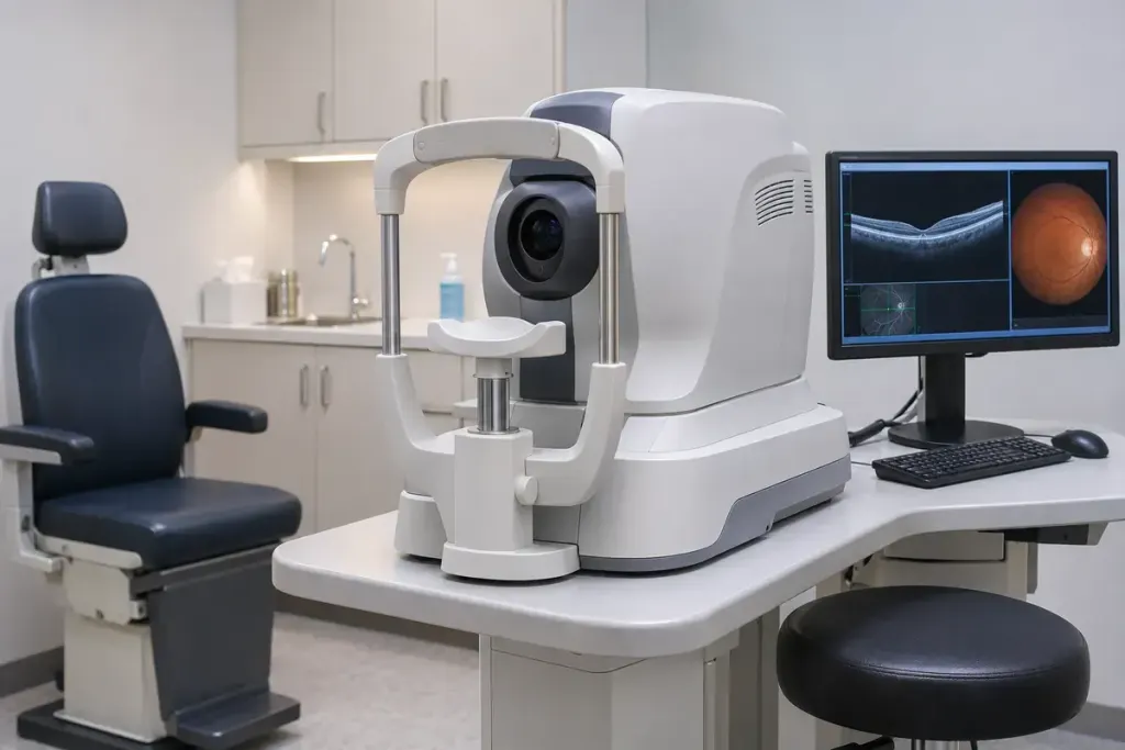
An eye scan report can show coloured bands, thickness maps and curved layers that look nothing like a familiar photograph of the back of the eye. Optical coherence tomography, or OCT, uses light to build cross-sectional images of tissue. It helps a clinician examine structures that a surface view alone does not describe in the same way.
The American Academy of Ophthalmology’s patient guide explains that OCT can map retinal layers and the optic nerve fibre layer. Measurements support assessment and follow-up for conditions such as glaucoma and retinal disease. A scan is interpreted alongside the rest of the examination; it is not a diagnosis you can reliably make from a coloured screenshot.
What the appointment may involve
You usually sit at the instrument with your head supported while it scans without touching the eye. The clinic may or may not use dilating drops. Ask beforehand whether dilation is expected and what that means for your journey home, especially if you planned to drive.
If holding a position is difficult, tell the clinic when arranging the appointment. Explain the practical difficulty—neck movement, tremor or another access need—so the team can discuss what is possible. You do not need to guess whether the machine can accommodate you from an online photograph.
Questions that make the result more useful
- Which structure was this scan intended to assess?
- Is this a first measurement or a comparison with an earlier scan?
- Was the image quality sufficient for the intended interpretation?
- What does the result change about the next step in my care?
Bring previous reports if the clinic requests them, and ask whether the original images are needed as well. A written number and a complete scan are different pieces of information.
New sudden loss of vision or severe eye symptoms should not wait for a routine imaging appointment; seek urgent medical advice. For planned care, the useful outcome is an explanation of what was measured and how it relates to your examination, rather than collecting a striking picture without context.